Home

Suspect investiție aproba cornea under microscope bunic Lacul Taupo Amestecat

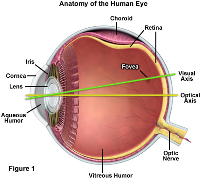
ZEISS Microscopy Online Campus | Microscopy Basics | Introduction

Cornea - an overview | ScienceDirect Topics

Transmission electron microscopy of normal cornea (A–D) and corneal... |  Download Scientific Diagram
Transmission electron microscopy of normal cornea (A–D) and corneal... | Download Scientific Diagram

Morphological evaluation of normal human corneal epithelium - Ehlers - 2010  - Acta Ophthalmologica - Wiley Online Library
Morphological evaluation of normal human corneal epithelium - Ehlers - 2010 - Acta Ophthalmologica - Wiley Online Library

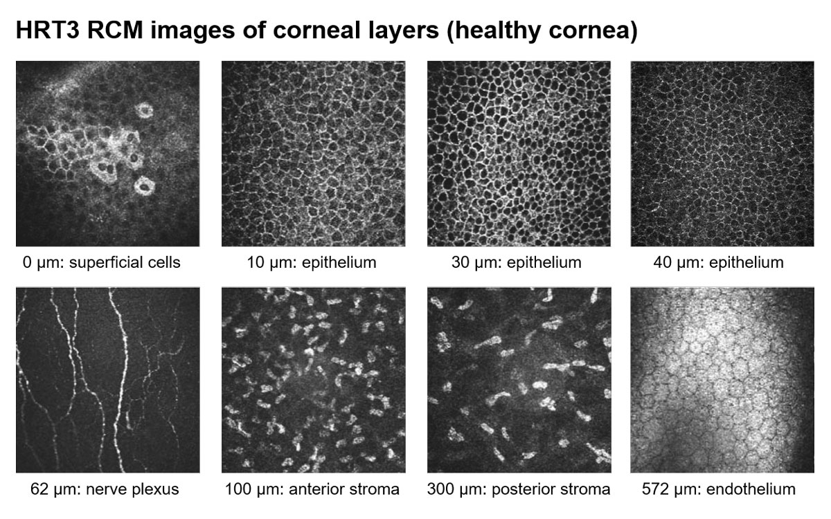
In Vivo Confocal Microscopy of the Cornea: New Developments in Image  Acquisition, Reconstruction, and Analysis Using the HRT-Rostock Corneal  Module. - Abstract - Europe PMC
In Vivo Confocal Microscopy of the Cornea: New Developments in Image Acquisition, Reconstruction, and Analysis Using the HRT-Rostock Corneal Module. - Abstract - Europe PMC
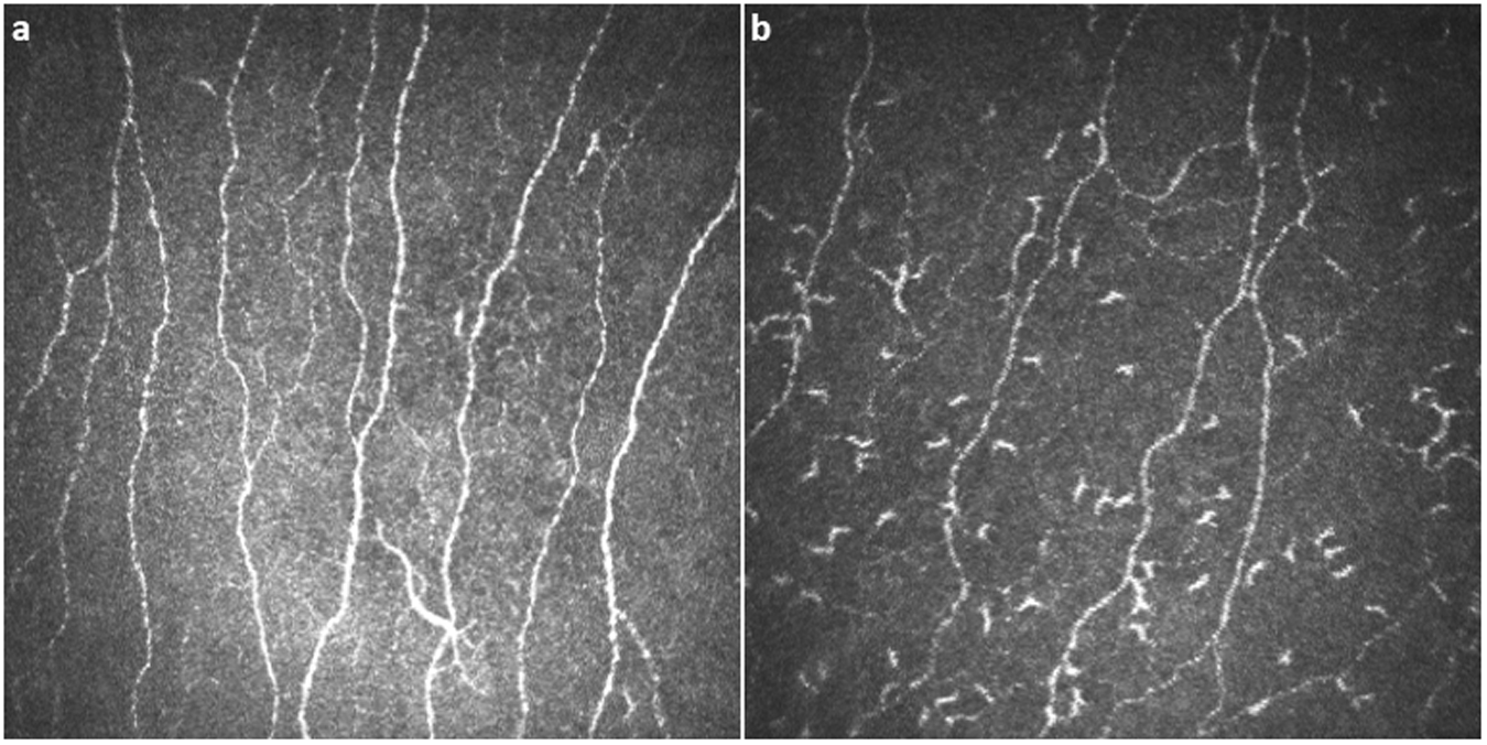
Corneal confocal microscopy detects corneal nerve damage and increased  dendritic cells in Fabry disease | Scientific Reports
Corneal confocal microscopy detects corneal nerve damage and increased dendritic cells in Fabry disease | Scientific Reports

PDF] ELECTRON MICROSCOPY OF THE HUMAN CORNEAL ENDOTHELIUM WITH REFERENCE TO  TRANSPORT MECHANISMS. | Semantic Scholar
PDF] ELECTRON MICROSCOPY OF THE HUMAN CORNEAL ENDOTHELIUM WITH REFERENCE TO TRANSPORT MECHANISMS. | Semantic Scholar

Light microscopy of control and KC corneas stained with PAS and Mayer's...  | Download Scientific Diagram
Light microscopy of control and KC corneas stained with PAS and Mayer's... | Download Scientific Diagram

Figure 1 from The normal and abnormal human corneal epithelial surface: a  scanning electron microscope study. | Semantic Scholar
Figure 1 from The normal and abnormal human corneal epithelial surface: a scanning electron microscope study. | Semantic Scholar
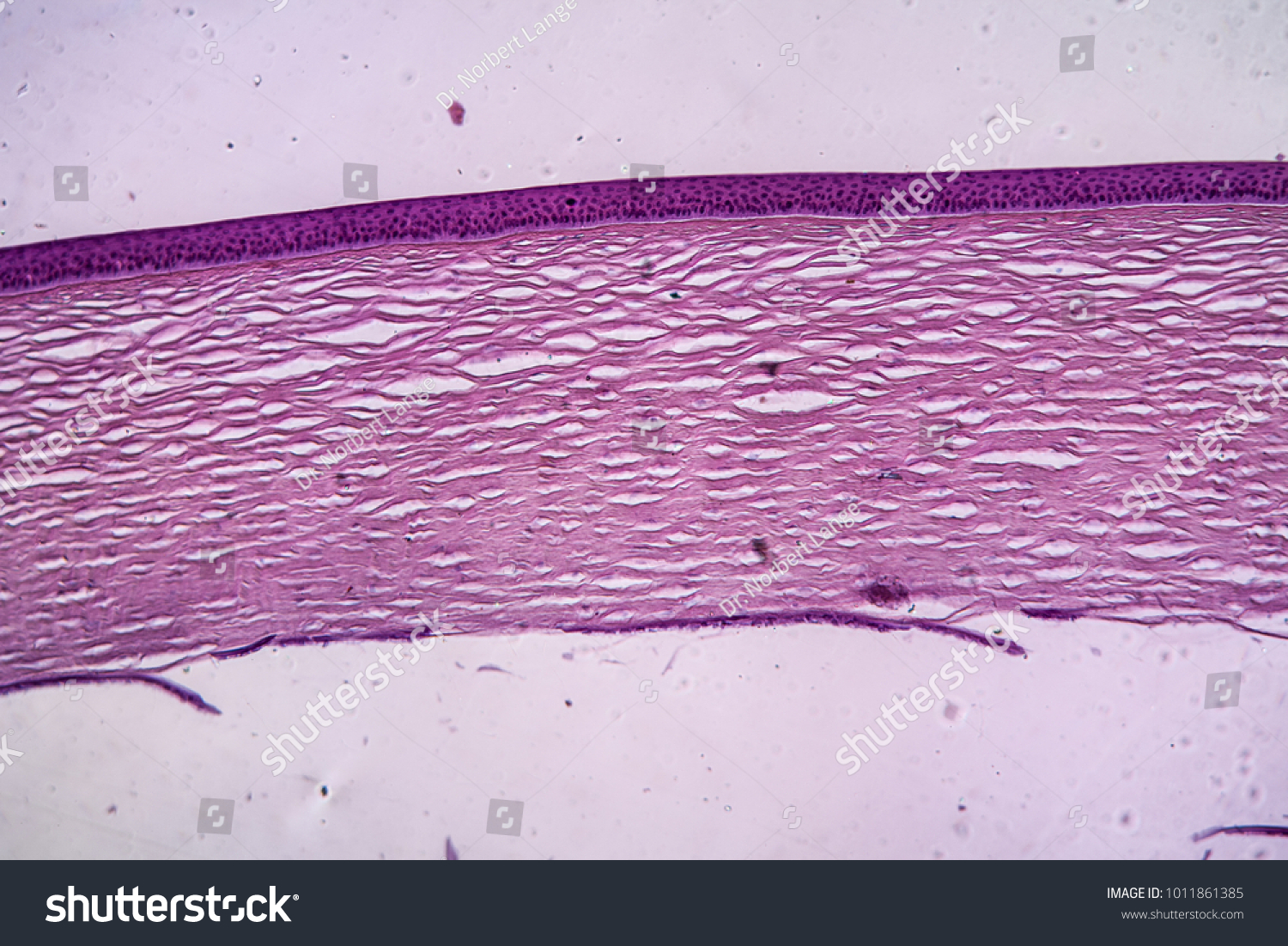
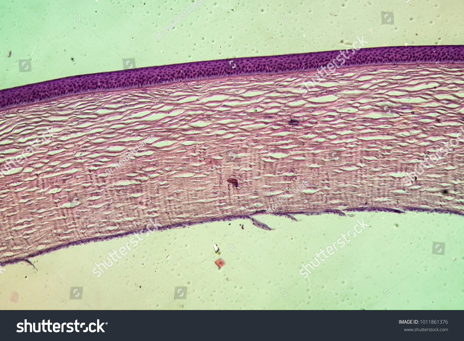
Cornea Eye Tissue Under Microscope 100x Stock Photo 1011861385 |  Shutterstock
Cornea Eye Tissue Under Microscope 100x Stock Photo 1011861385 | Shutterstock

Scanning Electron Microscopy of the Human Cornea

Human Structure Virtual Microscopy

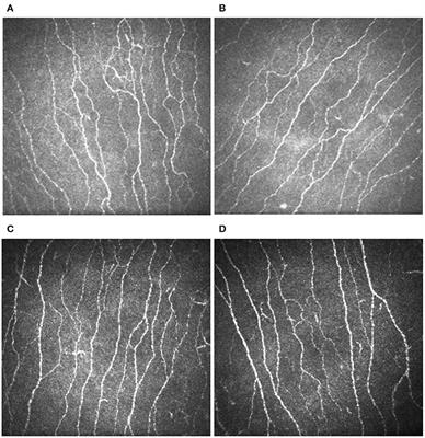
Frontiers | Corneal Confocal Microscopy Demonstrates Corneal Nerve Loss in  Patients With Trigeminal Neuralgia
Frontiers | Corneal Confocal Microscopy Demonstrates Corneal Nerve Loss in Patients With Trigeminal Neuralgia

Auricularia cornea, under the microscope | showing just how … | Flickr

Corneal confocal microscopy detects small nerve fibre damage in patients  with painful diabetic neuropathy | Scientific Reports
Corneal confocal microscopy detects small nerve fibre damage in patients with painful diabetic neuropathy | Scientific Reports

Light microscopic images showing the structure of the cornea stained... |  Download Scientific Diagram
Light microscopic images showing the structure of the cornea stained... | Download Scientific Diagram

Use Specular Microscopy to Diagnose Corneal Disease

In vivo confocal microscopy of the human cornea | British Journal of  Ophthalmology
In vivo confocal microscopy of the human cornea | British Journal of Ophthalmology

Scanning Electron Microscopy of the Human Cornea

Confocal microscopy in cornea guttata and Fuchs' endothelial dystrophy |  British Journal of Ophthalmology
Confocal microscopy in cornea guttata and Fuchs' endothelial dystrophy | British Journal of Ophthalmology

Cornea, Section Microscope Slide - Walmart.com

PDF] Scanning electron microscopy of the corneal endothelium of ostrich |  Semantic Scholar
PDF] Scanning electron microscopy of the corneal endothelium of ostrich | Semantic Scholar

Heidelberg Engineering brings back in vivo corneal microscope | Heidelberg  Engineering GmbH
Heidelberg Engineering brings back in vivo corneal microscope | Heidelberg Engineering GmbH

15 Normal axial cornea microscopic anatomy of commonly use species in... |  Download Scientific Diagram
15 Normal axial cornea microscopic anatomy of commonly use species in... | Download Scientific Diagram
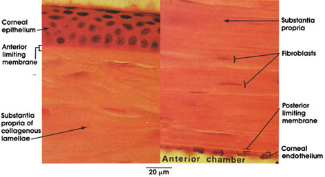
Histology Laboratory Manual

Cornea Eye Tissue Under Microscope 100x Stock Photo 1011861385 |  Shutterstock
Cornea Eye Tissue Under Microscope 100x Stock Photo 1011861385 | Shutterstock

Anatomy Atlases: Atlas of Microscopic Anatomy: Section 1 - Cells