Home

spate Prezice unpleasantly human cheak cell microscope lobby reţetă resturi

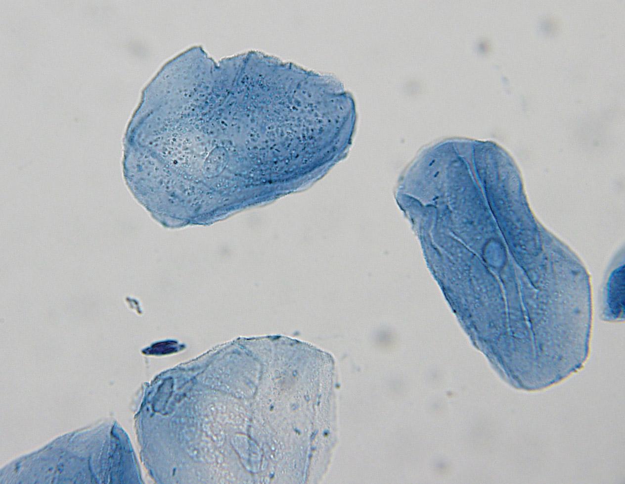
File:Human cheek cell.jpg - Wikimedia Commons

Cheek Cells
Cheek Cells

United Scientific Supplies 500-7"Human Cheek Cells" Prepared Microscope  Slide: Amazon.com: Industrial & Scientific
United Scientific Supplies 500-7"Human Cheek Cells" Prepared Microscope Slide: Amazon.com: Industrial & Scientific

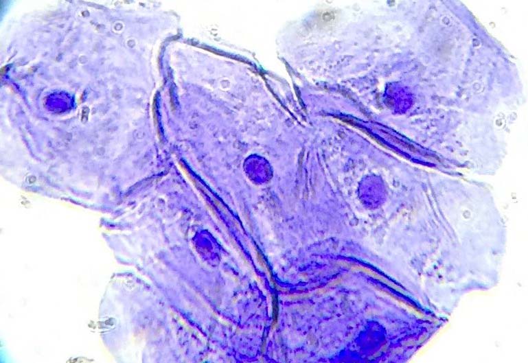
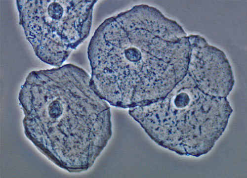
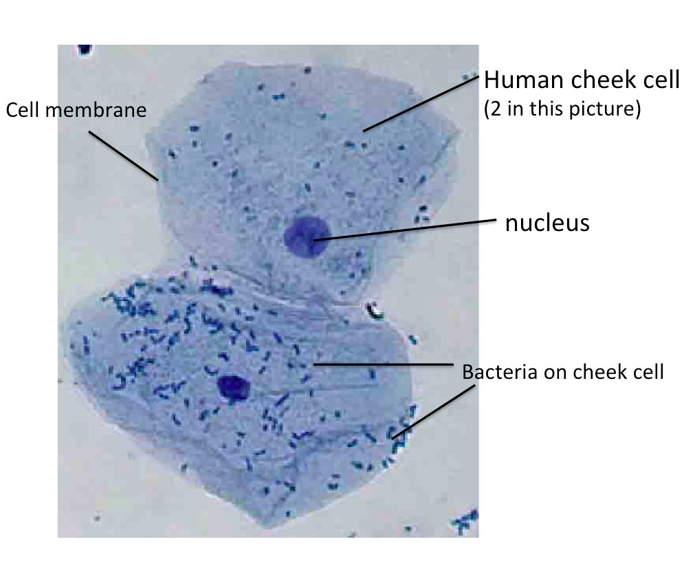
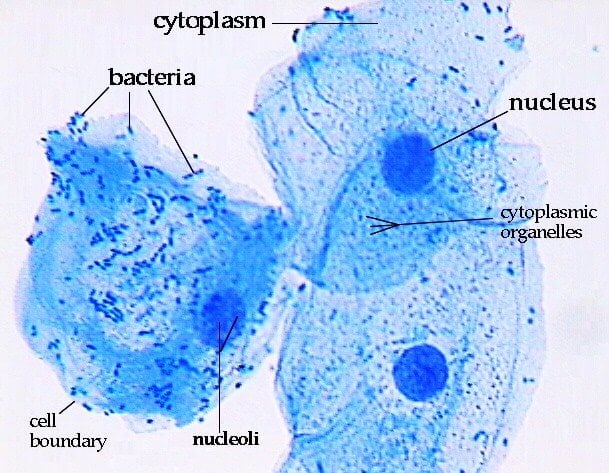
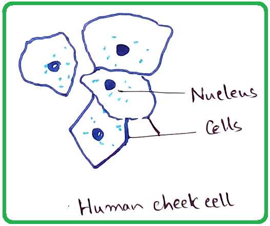
Identifying features of human cheek cells under microscope includes:

Looking at cheek cells under a microscope. | Fun science, Human body,  Things under a microscope
Looking at cheek cells under a microscope. | Fun science, Human body, Things under a microscope

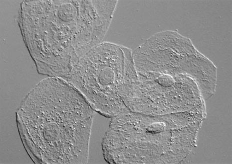
Buccal Epithelial Cells | Nikon's MicroscopyU

Solved Using this table from the Size Estimation module, | Chegg.com

Microscopic Image Of Human Cheek Cells Dyed Yellow Stock Photo - Download  Image Now - Bacterium, Biological Cell, Biopsy - iStock
Microscopic Image Of Human Cheek Cells Dyed Yellow Stock Photo - Download Image Now - Bacterium, Biological Cell, Biopsy - iStock

Human Cheek Epithelial Cells. the Tissue that Lines the Inside of the Mouth  is Known As the Basal Mucosa and is Composed of Stock Image - Image of  epithelial, diagnosis: 167106771
Human Cheek Epithelial Cells. the Tissue that Lines the Inside of the Mouth is Known As the Basal Mucosa and is Composed of Stock Image - Image of epithelial, diagnosis: 167106771

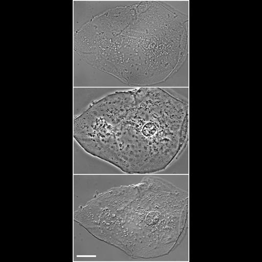
CIL:12594, Homo sapiens, squamous epithelial cell. CIL. Dataset

Amazing 27 Things Under The Microscope With Diagrams

Cheek cells 400X: General Biology Lab II: Loyola University Chicago

Human Cheek Epithelial Cells | Olympus LS

Introduction – Biology

Draw three types of cells (Cheek cell, Red blood cell, Elodea). Make sure  that you have also labeled these drawings with the cell structures that you  can see (nucleus, cell membrane, cell
Draw three types of cells (Cheek cell, Red blood cell, Elodea). Make sure that you have also labeled these drawings with the cell structures that you can see (nucleus, cell membrane, cell

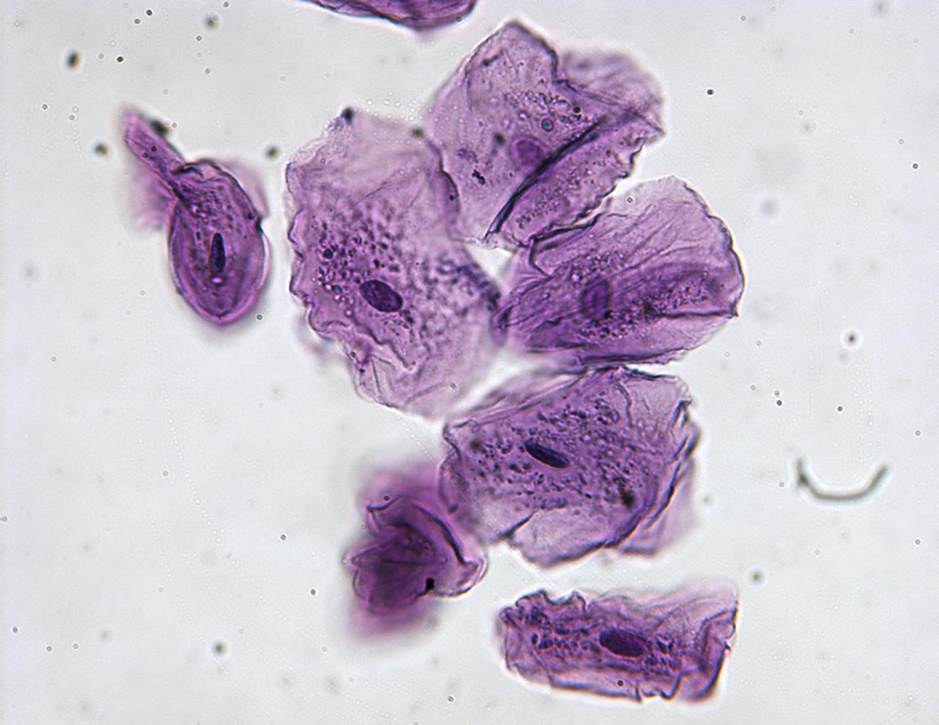
Human Cheek Cells Under the Microscope | Haematoxylin | Cell Membrane

Human cheek cell on PCS. (a) Bright field microscopy. (b) Phase... |  Download Scientific Diagram
Human cheek cell on PCS. (a) Bright field microscopy. (b) Phase... | Download Scientific Diagram

Hands-On Activity: Human Cheek Cells | Science Supply

Images of human epithelial cheek cells. (a) Image recorded by a... |  Download Scientific Diagram
Images of human epithelial cheek cells. (a) Image recorded by a... | Download Scientific Diagram

Mitochondria in human cheek cells - UWDC - UW-Madison Libraries

A human cheek cell and a spongy mesophyll cell are examined under a  microscope. Which structure are seen in both cells? - Quora
A human cheek cell and a spongy mesophyll cell are examined under a microscope. Which structure are seen in both cells? - Quora

Human Cheek Cell Station